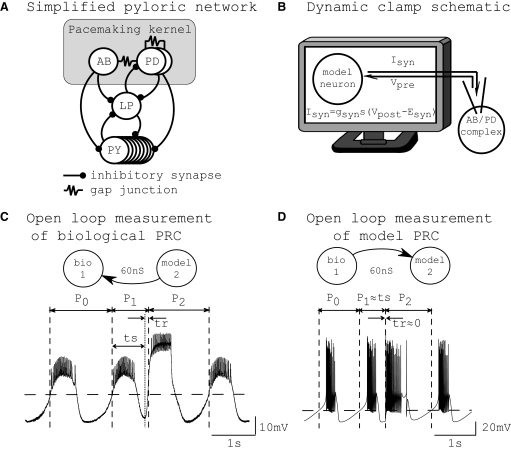
FIG. 1.
Experimental setup. A: simplified pyloric network. The pacemaking kernel (anterior burster/pyloric dilator [AB/PD] complex) consists of the AB and PD neurons, which are coupled via gap junctions. B: schematic of the dynamic clamp. The membrane voltage recorded in the AB/PD complex was used to calculate synaptic current (Isyn) received by the model cell. The model cell was updated in real time and the updated model neuron's membrane voltage was used to calculate Isyn received by the AB/PD complex. C: measurement of phase resetting in a biological neuron. Shown is a membrane potential recording from a free-running AB/PD complex with intrinsic period P0, perturbed at ts (dotted vertical line) by an excitatory synaptic input, gsyn = 60 nS and duration = 650 ms. An upward crossing of the voltage threshold (horizontal dashed line; see Phase response curves) was defined as the start of a burst and assigned a phase of zero. The first burst after the perturbation was advanced such that the first perturbed period P1 was <P0. The subsequent period P2 was delayed by a small amount such that P2 > P0. In this case, a new burst was triggered after a short delay of 100 ms, so that P1 > ts and tr = 100 ms. D: measurement of phase resetting in a model neuron. Analogous to the biological neuron, the free-running model neuron was perturbed at ts by an excitatory synaptic input with gsyn = 60 nS. This perturbation was strong and triggered a new burst immediately, such that P1 ≈ ts and tr ≈ 0. The burst threshold is shown as a horizontal dashed line. (B adapted from DailyClipArt.net.)

