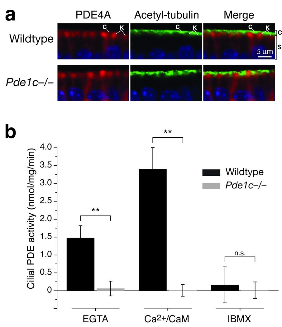
Figure 3. Knockout of PDE1C eliminates all PDE activity from OSN cilia.
(a) Co-immunostaining for PDE4A (red) and the cilia marker acetylated tubulin (green) on sections of OE from wildtype and Pde1c−/− mice. Shown are apical portions of the OE under high magnification. In both wildtype and Pde1c−/− mice, immunoreactivity for PDE4A and acetylated tubulin appears in distinct spatial domains. C, cilial layer; K, dendritic knobs; S, supporting cell layer. Sections are counterstained with DAPI. Scale bar: 5 µm. (b) PDE catalytic activity assay in cilia preparations from wildtype and Pde1c−/− mice. PDE activity in wildtype cilia was increased by addition of Ca2+/CaM and inhibited by addition of IBMX. Activity from Pde1c−/− cilia under all conditions was virtually undetectable, similar to wildtype cilia treated with IBMX. Error bars are 95% CI. Data for each genotype are the average of 5 independent preparations (mice). Each preparation was assayed in duplicate.

