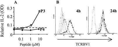
Figure 3.
Engagement with the MHC:peptide complex down-regulates the TCR. TCR37.33 were incubated for 24 h with equal numbers of APC prepulsed with or without increasing concentration of peptides. (A) At each peptide concentration, the supernatants were collected after 24 h and the released IL-2 assessed by ELISA. Challenge with peptide P3 is represented by the open symbols, whereas peptide P4 is represented by the filled symbols. APC with the irrelevant MBP peptide at 10 μM did not induce any IL-2 release (broken line). (B) After 4 and 24 h, cells were stained for expression of human TCRBV1 and fixed in 1% formaldehyde. TCR37.33 cells were tightly gated on forward scatter/side scatter to exclude the APC. TCR expression was tested in TCR37.33 incubated with APC and the irrelevant MBP peptide 13–32 (solid line, mean fluorescence intensity 97.8), peptide P3 (dotted line, mean fluorescence intensity 8.76) or peptide P4 (dashed line, mean fluorescence intensity 12.19). Peptides were used at 10 μM. The gray line represents TCR37.33 stained with a rat anti human isotype control (mean fluorescence intensity 5.8). The data are representative of at least four different experiments.

