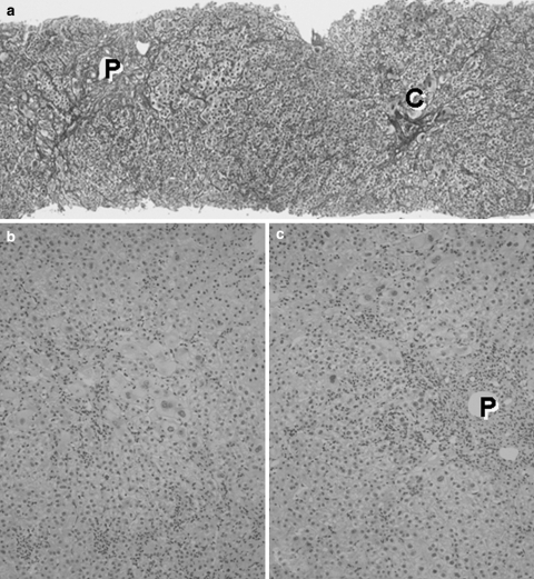
Fig. 3.
Histologic picture of the liver biopsy specimen in de novo acute hepatitis B. (a) Hepatic lobular architecture is preserved and fibrous thickening of the walls of terminal hepatic venules (C) is seen. Portal triads (P) are not fibrously enlarged. The regular trabecular pattern of hepatocytes is slightly disrupted. Silver impregnation, 10×. (b) In hepatic lobules, anisocytosis of hepatocytes is conspicuous and many areas of focal necroses are seen. Inflammatory mononuclear cells are markedly infiltrated into hepatic sinusoids. Hematoxylin-eosin stain, 25×. (c). Portal triads are not fibrously enlarged, indicating an acute nature of this disease condition. Focal necroses are also conspicuous around portal triads. Hematoxylin-eosin stain, 25×

