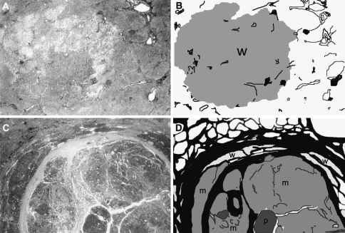
Fig. 1.
A typical early hepatocellular carcinoma (HCC) with indistinct (vague) margin and a typical advanced HCC with distinct margin. a A panoramic view of an early HCC of 1.4-cm diameter. b An illustration of panel a. The tumor margin is indistinct. The cancer tissue consists only of well-differentiated HCC (w). Pre-existing hepatic architectures (portal tracts) are well preserved within the nodule. The noncancerous tissue is precirrhotic (not advanced cirrhosis). c A panoramic view of an advanced HCC of 2.6-cm diameter. d An illustration of panel c. The tumor margin is distinct with thick fibrous capsule. The cancer tissue consists of well- (w), moderately (m), and poorly (p) differentiated types. Well-differentiated type tissue is found only in the peripheral area of the nodule. The cancer nodule is occupied mostly by moderately and poorly differentiated types. The noncancerous tissue corresponds to advanced cirrhosis. Adapted from Kondo and Ebara [7]

