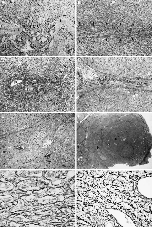
Fig. 3.
Stromal invasion of hepatocellular carcinoma (HCC). a Crossing type. Cancer tissue (HCC) invades across fibrous septa (f) of tumor nodule. b Longitudinal type. Tumor cells grow longitudinally within fibrous septa (arrowheads). c Irregular type. Portal areas are irregularly invaded by tumor cells. Portal veins (p), an artery (a), and a bile duct (b) are surrounded by tumor cells. Arrowheads show longitudinal type invasion in the adjacent fibrous septa. d A noncancerous area without invasion. A portal area and fibrous septa are clearly seen. e Pseudoinvasion. Benign noncancerous cells are found in the fibrous stroma. f A panoramic view of stromal invasion. In the noncancerous area without invasion [the area of (a)], the fibrous septa are clearly seen. In the area of tumor spread [the area of (b)], the septa are indistinct because stromal invasion of the longitudinal and irregular types (b, c) reduced the amount of the fibrous component. In the area of (c), hepatic architecture is severely disturbed because of stromal invasion of crossing type. g Silver staining of the pseudoinvasion. The liver cells are clearly surrounded by reticulin fibers. h Silver staining of the true invasion. Carcinoma cells are not surrounded by reticulin fibers. Adapted from Kondo et al. [8, 14]

