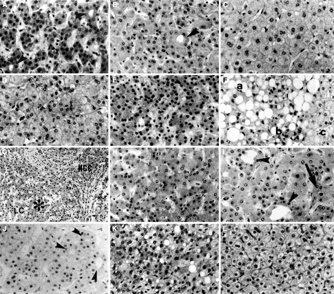
Fig. 4.
Various histological findings useful for the biopsy diagnosis of hepatocellular carcinoma. a A well-differentiated hepatocellular carcinoma (HCC). Arrowheads show microacinar formation. b A dysplastic nodule. An arrowhead shows microacinar formation. c Noncancerous tissue. d A very well-differentiated HCC. Although it does not show hypercellularity, it shows stromal invasion outside this figure (same case as in Fig. 3b, c). Noncancerous tissue of this case shows lower cellularity than this HCC. e A large regenerative nodule with hypercellularity. In some cases of cirrhosis, ordinary regenerative nodules show such high cellularity. f An HCC with fatty change. In the area (a) with fatty change, diagnosis is difficult because true cellularity is secondarily reduced by fatty change. In the area of (b) without fatty change, cellularity is clearly higher than in the noncancerous area (c). g and h An area with a mixture of HCC cells and benign cells. g Low magnification. (HCC) shows cancer area with stromal invasion. (LC) shows noncancerous liver cirrhosis. In the area of (*), cancer cells and benign cells are mixed. h High magnification of the (*) area. It shows intermediate cellularity between well-differentiated HCC (a) and noncancerous tissue (c). i Liver tissue near a metastatic tumor. The arrow shows bile. Microacinar formations (arrowheads) are thought to be caused by cholestasis. j Nodular regenerative hyperplasia. Hypercellularity and microacinar formation (arrowheads) make this tissue resemble well-differentiated HCC. k Focal nodular hyperplasia (FNH) with high cellularity. It resembles well-differentiated HCC. l FNH without hypercellularity. It is similar to ordinary noncancerous tissue (HE stain). Adapted from Kondo et al. [8]

