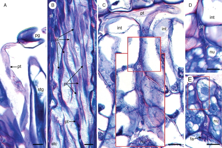
Fig. 4.

Pollen tube growth in Tofieldia glutinosa. (A) Pollen tube growing out of a pollen grain on the stigma. (B) Numerous pollen tubes grow through the stylar canal. (C) Pollen tube growth through the micropyle. This image is a composite of three serial histological sections of the same ovule. Red boxes indicate digital superposition of pollen tube and surrounding tissue from adjacent histological sections. (D) Penetration of the nucellus by a pollen tube. Pollen tube growth through the nucellus is intercellular. (E) Pollen tube growth through the nucellus and into the filiform apparatus of the female gametophyte. The red box indicates digital superposition of the pollen tube and surrounding tissue from the adjacent histological section. All sections were stained with toluidine blue. fa, Filiform apparatus; int, integument; nu, nucellus; pg, pollen grain; pt, pollen tube; st, style; stc, stylar canal; stg, stigma. Scale bars = 10 µm.
