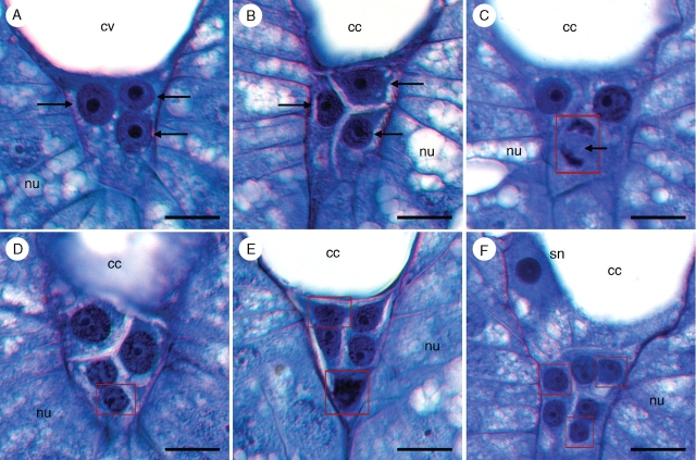
Fig. 9.
Proliferation of antipodal nuclei in Tofieldia glutinosa. (A) Three antipodal nuclei (arrows) prior to cellularization of the female gametophyte. (B) Three uninucleate antipodal cells (arrows) following cellularization of the female gametophyte. (C) Antipodal nucleus in mitosis (arrow). Mitotic divisions of the antipodals are usually asynchronous. (D) Single binucleate antipodal and two uninucleate antipodals. (E) Two binucleate antipodals and a single uninucleate antipodal. (F) Three binucleate antipodal cells. The secondary nucleus is also visible in this section. All sections were stained with toluidine blue. Red boxes indicate digital superposition of nuclei from adjacent histological sections. cc, Central cell; cv, central vacuole; nu, nucellus; sn, secondary nucleus. Scale bars = 10 µm.

