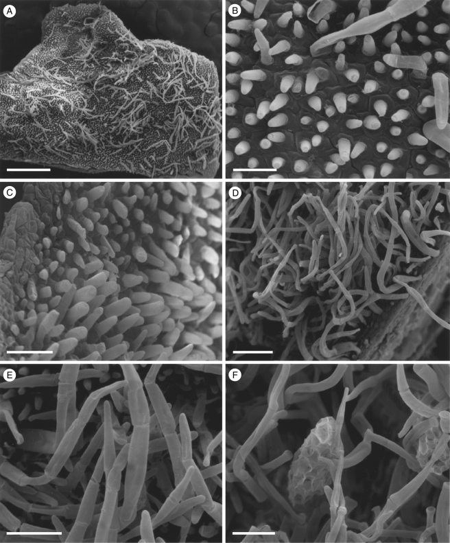
Fig. 2.
Labellar micromorphology of Scuticaria. Detail of labellar surface (A–F) of S. adverna (S. bahiensis K.L. Davies & M. Stpiczyńska; accession no. K57975) showing uniseriate, multicellular trichomes (A, D–F), conical papillae (B, C) and multicellular, scale-like structures (F). Scale bars = 900, 80, 90, 200, 100 and 100 µm, respectively.

