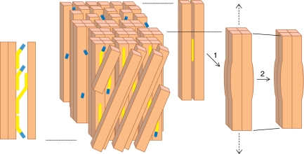
Fig. 3.
Model of the G-layer and its attachment to the S2 layer, depicting the proposed roles of xyloglucan (yellow), XTH with XET activity (blue) and cellulose fibrils (beige) in the development of tensile stress during maturation. The G-layer is shown from outside through two layers of S2 cellulose microfibrils orientated at an angle. The G-layer has several layers of cellulose microfibrils orientated axially, which aggregate during maturation to form a lattice-like structure (not pictured here) locally forming groups of four microfibrils that could trap short xyloglucan chains inside, as shown in 1. This induces the longitudinal tensile stress that leads to macrofibril shortening, as shown in 2. Xyloglucan makes cross-links between G and S2 layers as shown on the left. The XTH enzyme saturates the cut ends of xyloglucan ready to reconnect them to a suitable acceptor. Mechanical rupture of a cross-link would thus be rapidly repaired by transglucosylation. Xyloglucan-free XTH enzyme is stored in the G-layer. For more details see the text.

