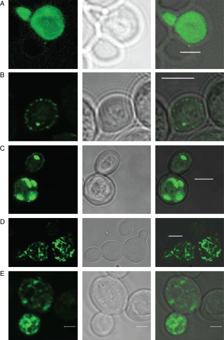
Fig. 6.
Localization of MbNRAMP1–GFP in yeast. Upper case letters show different GFP staining patterns: left, GFP; middle, differential interference contrast (DIC) of the same fields; right, GFP with DIC superimposed. (A) Free GFP staining as control in normal growth conditions; homogenous fluorescence. (B–E) Examples of MbNRAMP1–GFP localization patterns in yeast. MbNRAMP1 exhibits a shift in cellular localization with the iron status. (B) MbNRAMP1–GFP staining in peripheral vesicles, under normal growth conditions. (C) MbNRAMP1–GFP is localized in brightly fluorescent intracellular vesicles seen as irregular structures, under normal growth conditions. (D) MbNRAMP1–GFP staining in the whole cell in the presence of BPDS and EGTA. (E) MbNRAMP1–GFP staining in the whole cell, and unequally accumulated on the vacuole-like structure surface in the presence of BPDS and EGTA. Scale bars: (A–D) = 4 µm; (E) = 2 µm.

