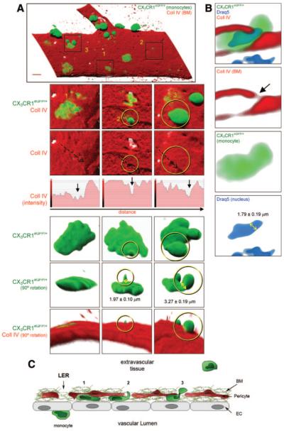
Figure 3.
Monocyte protrusion formation while penetrating LERs. A, Monocytes embedded within the BM and exhibiting at least 3 distinct morphological shapes after CCL2-stimulation. B, Venular cross-section showing a monocyte squeezing both its body and nucleus. C, Schematic diagram of the different stages of monocyte migration. (Please see the supplemental materials for details).

