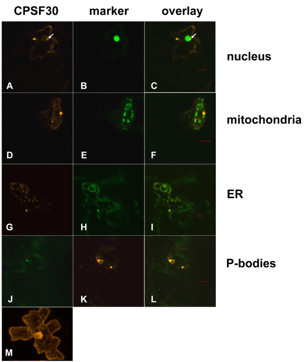
Figure 1.
Subcellular distribution of AtCPSF30 in tobacco cells. In this figure, the overlays of the two images corresponding to pairs of fusion proteins are shown in the column on the right (panels C, F, I, and L). The AtCPSF30 is visualized in panels A, D, G, and J. Markers used to assess the subcellular location of AtCPSF30 were: nuclear marker (GFP-AtZFP11), panel B; mitochondrial marker (GFP-NDA2), panel E; endoplasmic reticulum marker (the synthetic localization sequences carried in the mgfp4-ER plasmid described by Haseloff et al. [21]), panel H; and the P-body marker (Dcp2), panel K. The distribution of unmodified DSR is shown in panel M. Red bars are 10 μm size markers.

