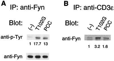
Figure 3.
Increased Fyn tyrosine phosphorylation and association with TCR/CD3 complex after antagonist stimulation. AD10 cells (1 × 10⁷) were stimulated with antagonist (T102G) pulsed, agonist (PCC 88–104) pulsed, or unpulsed DCEK.ICAM APCs (1 × 10⁷). After 1 min of stimulation, cells were lysed, immunoprecipitated with anti-Fyn or CD3ɛ antibody, and analyzed by SDS/PAGE. Proteins were transferred to membranes and immunoblotted with: (A) antiphosphotyrosine antibody (Upper) or (B) anti-Fyn. Membranes were subsequently stripped and analyzed for Fyn protein (A Lower). The numbers under Upper indicate the intensity of staining relative to that observed in the unstimulated controls.

