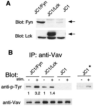
Figure 5.
Vav tyrosine phosphorylation in transfected JCaMl (JCl) cells. (A) An equal number (2.5 × 10⁵) of JCl/Fyn (Fyn+, Lck−) JCl/Lck (Lck+, Fynlo), or JCl cells were lysed in 1% Nonidet P-40 and analyzed by immunoblotting with either anti-Fyn (Upper) or anti-Lck antibodies (Lower). (B) JCl/Fyn, JCl/Lck, or JCl cells (1 × 10⁷) were incubated with or without 10 μg/ml anti-CD3 antibodies for 30 min on ice. After washing, cells were incubated with rabbit anti-mouse IgG antibody (20 μg/ml) at 37°C for 3 min. After washing with cold PBS, cells were lysed, immunoprecipitated with anti-Vav, and analyzed by SDS/PAGE. Proteins were transferred to membranes and immunoblotted with antiphosphotyrosine antibody. Films were exposed for 5 sec (Left Upper) or for 2 min (Right Upper JCl*); membranes were subsequently stripped and analyzed for Vav protein (Lower). Numbers indicate the fold increase in Vav phosphorylation after TCR crosslinking relative to the Vav phosphorylation in unstimulated cells.

