
Figure 1. CCN1 is expressed in prostate cells and supports prostatic cell adhesion through integrins.
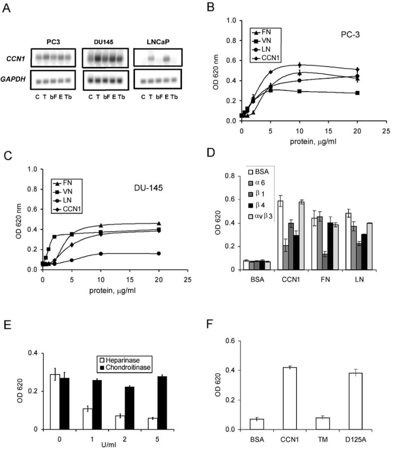
A. RNA blot of total RNA from PC-3, DU145, and LNCaP cells treated with TPA (T; 10 nM), bFGF (bF; 10 ng/ml), EGF (E; 3 ng/ml), or TGF-β (Tb; 10 ng/ml) for 1 hr, hybridized to cDNA probes for CCN1 and GAPDH. B. PC-3 cells adhered on plates coated with various concentrations of CCN1, FN, VN, and LN for 30 min. Adherent cells were stained with methylene blue and extracted dye was quantified by absorbance at 620 nm. C. Adhesion of DU145 cells on various substrates as described above. D. PC-3 cells were pre-treated with antibodies against α6, β1, β4, or αvβ3, integrins before adhering to plates coated with indicated substrates and cell adhesion measured. E. PC3 cells were treated with 1-5 u/ml of heparinase or chondroitinase and their adhesion to CCN1-coated plates measured. F. PC-3 cell adhesion to plates coated with BSA, CCN1, or the CCN1 mutants TM (α6β1-HSPG binding-defective) and D125A (αv binding-defective) measured by methylene blue staining.