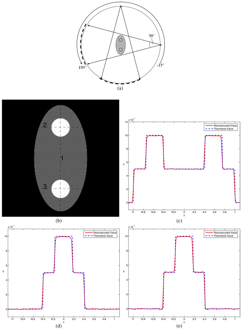
Figure 3.
Simulation for case 1. Part (a) shows the diagram for the scan configuration, in which the view angle range equals 180° plus the fan angle 30°. The shaded area shows the part of the object which can be accurately reconstructed based on the data-sufficiency condition. Part (b) is the reconstructed image, and parts (c)–(e) are the profiles for the three lines in (b), respectively. The entire phantom is accurately reconstructed.

