
Figure 1.
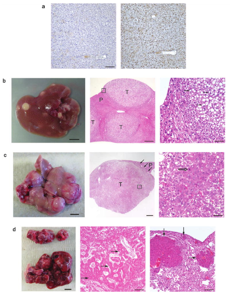
Accelerated tumorigenesis in p53-deficient livers compared with wild-type livers. (a) Albumin-Cre (Alb-Cre) efficiently deletes the floxed-stop (lsl) cassette within the Rosa26-lsl-SB11 transgene, allowing for SB transposase expression and subsequent somatic transposition events. Left panel, immunohistochemistry (IHC) of (Alb-Cre; Rosa26-lsl-SB11) double-transgenic liver section treated without the primary antibody (negative control). Right panel, IHC of serial liver section treated with the primary anti-SB transposase antibody. Sections were lightly stained with hematoxylin after IHC. Scale bar, 100 μm. (b) Quadruple male transgenic mouse liver at 159-days, displaying many preneoplastic nodules (left panel, scale bar, 0.5 cm). Middle (low magnification) and right (high magnification of boxed area from middle panel) panels show the tumor histology of large preneoplastic nodules using hematoxylin-eosin (HE) staining. These proliferative lesions were often compressing surrounding parenchyma and the cells within the preneoplastic foci and adenomas were frequently vacuolated, containing distinct lipid vacuoles or clear cytoplasm (arrows). Nuclei were equal to or smaller in size than those in the normal hepatic parenchyma, and occasionally contained mitotic figures indicative of cell division. Proliferative lesions were frequently bordered by hepatocytes with markedly enlarged nuclei that were occasionally karyomegalic. T, preneoplastic tumor nodule; P, parenchymal liver cells; scale bars for middle and right panels were 500 μm and 100 μm, respectively. (c) Triple-transgenic male experimental mouse liver at 330-days showing advanced tumor development. Note the many large irregular nodules showing a hypervascular phenotype (left panel, scale bar, 0.5 cm). Middle panel shows the HE histological section of one large neoplastic nodule typical of hepatocellular adenoma consisting of variably vacuolated hepatocytes filled with lipid. Three black arrows indicate border between adenoma and non-neoplastic hepatic parenchyma (P), which is slightly compressed. Right panel shows high magnification of boxed area in the middle panel. Note enlarged nuclei of hepatocytes with moderate variation in nuclear size, prominent nucleoli, and mitotic figure (open arrow). T, preneoplastic tumor nodule; P, parenchymal liver cells; scale bars for middle and right panels were 1000 μm and 50 μm, respectively. (d) Triple-transgenic male experimental mouse at 440-days with HCC (bottom, left panel) and lung metastasis (top, left panel). HE staining of the liver (middle panel) and lung (right panel), showing advanced HCC in the liver and its metastases into the lung. A partial HCC section composed of irregular trabeculae of neoplastic, diffusely necrotic hepatocytes (black arrows) that are multifocally vacuolated. Trabeculae are separated by dilated sinusoids containing variable amount of fibrin. The lung contains multiple variably sized metastatic nodules of HCC (black arrows) that markedly compress the pulmonary parenchyma. Pulmonary alveoli are filled with large numbers of foamy macrophages. Scale bar, 100 μm.