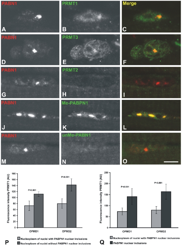
Figure 2. PRMT1 and PRMT3 accumulate in OPMD intranuclear inclusions.
(A–O) Immunofluorescence was performed on biopsy samples of quadriceps muscle from two OPMD patients using the indicated antibodies. Scale bar = 5 µm. (P–Q) From each patient (OPMD1 and OPMD2), approximately 60 nuclei labelled with anti-PRMT1 antibody were analyzed. The mean fluorescence intensity measured in the nucleoplasm is higher in nuclei devoid of inclusions compared to nuclei with inclusions (P). The mean fluorescence intensity measured in the nuclear inclusions is approximately two-fold higher than the intensity measured in the nucleoplasm of nuclei with inclusions (Q). Quantitative measurements of fluorescence intensity were performed with Image J software and expressed in arbitrary units (AU). Results are presented as mean fluorescence intensity±SE; p values (Student's t-test) are indicated.

