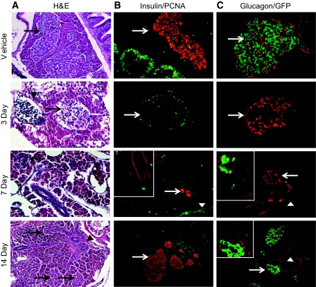
FIG. 3.
Regeneration of zebrafish islets after STZ treatment. A: Hematoxylin and eosin (H&E) staining of paraffin sections at 3, 7, and 14 days. Vehicle and 14 day STZ: 200× magnification; 3 day and 7 day: 400× magnification. Arrows: islets. Arrowheads: blood vessels. B: STZ Ins/PCNA: insulin antibodies (visualized with red fluorescent secondary antibodies) mark β-cells. PCNA+ dividing cells are green. Arrows identify islets. Arrowheads: ducts; vehicle: numerous dividing PCNA+ cells are located at the base of intestinal villi. A few non–insulin expressing dividing cells are scattered throughout the islet and surrounding exocrine pancreas (400× magnification); 3 day STZ: a mantle of PCNA-positive cells surrounds affected islets (400×); 7 day STZ: dividing cells are located in and around ducts (400× magnification); inset (200× magnification): CK18 (red) labeling of ducts. Insulin+ cells are green; 14 day STZ: 200× magnification. Large islets with scattered dividing cells have appearance similar to vehicle-injected zebrafish. Dividing cells surround insulin-negative areas, similar to the PCNA expression observed after 3 days. C: STZ glucagon/GFP; vehicle (400× magnification): β-cells (green) and α-cells (red). Arrow: islet; 3 day STZ: glucagon+ cells within islet remnant (400×); 7 day STZ: glucagon labels GFP-negative islet attached to duct (200×). Inset (400×): glucagon (red) outlines islets ± β-cells (green); 14 day STZ: ductal hyperplasia (200×) with glucagon staining (red). Inset (600×): the ductal epithelium is continuous with the islet (confocal image). (A high-quality digital representation of this figure is available in the online issue.)

