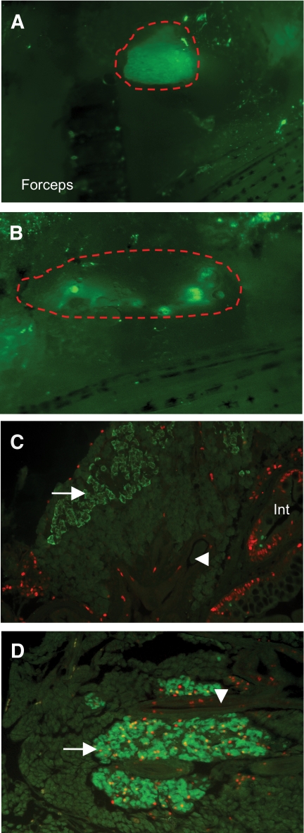
FIG. 5.
Regeneration after pancreatectomy. A and B: Right side of intact, living zebrafish before (sham, A) and 14 days after (Ptx, B) surgical removal of the GFP+ pancreas (red outline). The tip of the forceps used to remove the pancreas is visible (100× magnification). C: Paraffin section of sham-operated pancreas with few PCNA+ dividing cells (PCNA, red) except in the intestine (Int). β-Cells are green (arrow; 200× magnification). D: Many red PCNA+ dividing cells in ducts (arrowhead) and in nuclei of regenerating β-cells (yellow; 200× magnification). (A high-quality digital representation of this figure is available on the online issue.)

