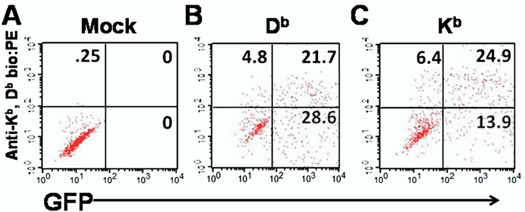
Fig. 2.
Flow cytometry analysis of efficient class I and green fluorescent protein (GFP) co-transfection in Cos-7 cells. Co-transfected Cos-7 cells were stained with antibody that binds both the Db and Kb class I molecule. Shown are a representative well of (A) cells transfected with empty pcDNA 3.1 His A vector, (B) Db plus GFP expression vectors, and (C) Kb plus GFP expression vectors. Note that cells within the upper right quadrant denotes successful co-expression of GFP and class I molecules.

