
Figure 8.
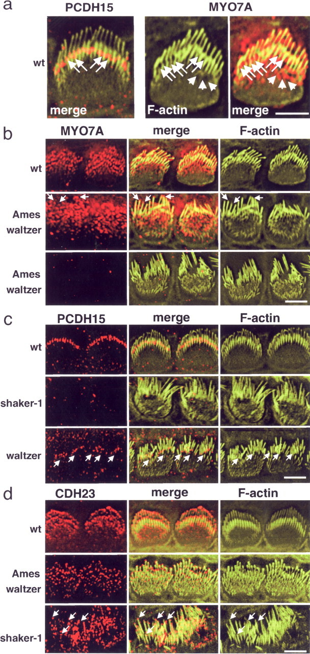
Localization of PCDH15 and MYO7A in wild-type (wt) and mutant mice. Inner hair cells of P5 animals are shown. a, The left panel shows cells from a wild-type mouse stained for PCDH15 (red) and F-actin (green; same cell as that shown in Fig. 1C). The middle and right panels show a cell stained for F-actin (green) and MYO7A (red). Note that two rows of stereocilia could be resolved. PCDH15 and MYO7A were localized toward the base of the longest stereocilia (arrows); MYO7A was also localized to the cuticular plate (arrowheads) and toward tips of stereocilia. b, MYO7A localization in PCDH15-deficient Ames waltzerav3J mice. In some hair cells, MYO7A could still be visualized, but it was mostly found at the apical hair cell surface and also at tips of stereocilia (arrows). In many hair cells, MYO7A could no longer be detected. Note that the wild-type control includes the hair cell shown at higher magnification in a. c, PCDH15 localization in MYO7A-deficient shaker-1 mice and CDH23-deficient waltzer mice. PCDH15 could no longer be detected in stereocilia in shaker-1 mice but was still confined toward the base of stereocilia in waltzer mice. Note the disorganization of the stereociliary bundles in the waltzer mice. d, CDH23 localization in PCDH15-deficient Ames waltzerav3J mice and MYO7A-deficient shaker-1 mice. CDH23 was still localized at the tips of stereocilia in the two mouse strains. Note that, in the Ames waltzerav3J mice, stereocilia were splayed but less affected than in shaker-1 mice. Because of the strong variations in stereociliary length in shaker-1 mice, it was more difficult to resolve CDH23 distribution, but it was clearly detectable at tips (arrows). Scale bars: a, 5 μm; b–d, 4 μm.