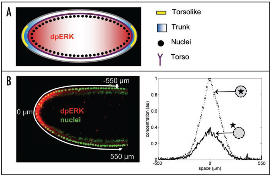
Figure 2.
(A) Torso receptors (purple) are uniformly distributed along the plasma membrane of the embryo. Inactive ligand (Trunk) is distributed uniformly in the extracellular matrix; it is converted into an active and diffusible form (blue) by Torsolike (yellow), a protein localized at the poles of the embryo.10 Torso-Trunk complex signals through the MAPK signaling cascade, which leads to MAPK phosphorylation. (B) Quantified pattern of MAPK phosphorylation. Left: fluorescent image of the anterior part of the embryo; nuclei are stained in green and phosphorylated MAPK is stained in red. Right: Gradients of nuclear and cytoplasmic phosphorylated MAPK. Reproduced from Berezhkovskii et al. PNAS 2009.

