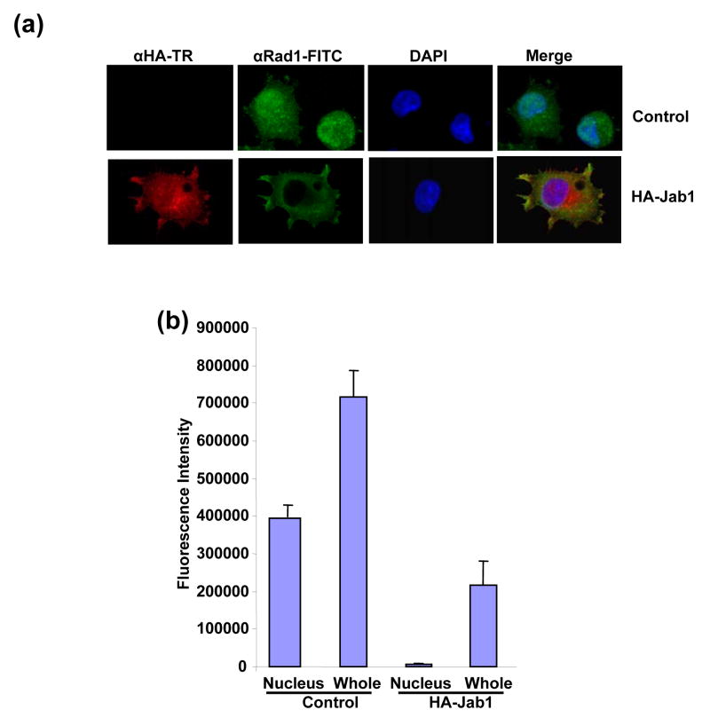
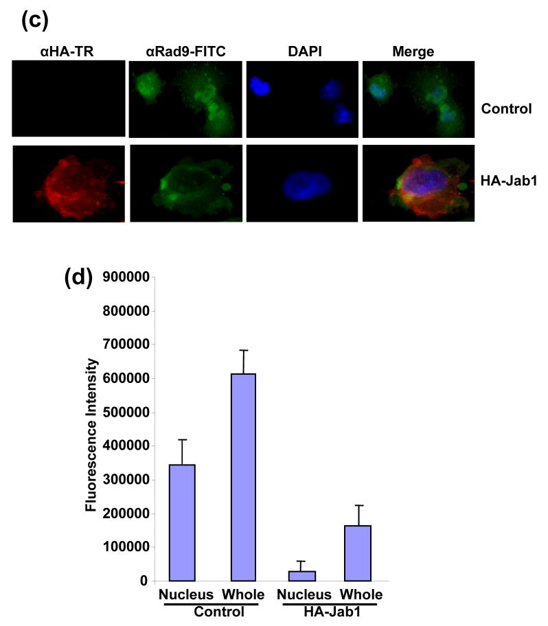
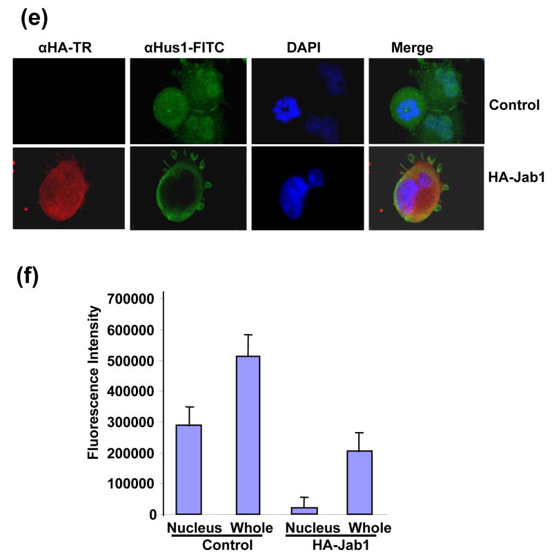
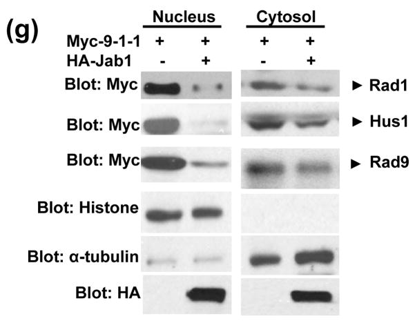
Figure 4. Subcellular localization of 9-1-1 in the presence and absence of Jab1.
(a, c and e) Jab1 translocates 9-1-1 proteins from nucleus to cytoplasm in PANC-1 cells analyzed by fluorescence colocalization. PANC-1 cells were infected with retrovirus containing empty vector (pSMCVneo, Control) or pSMCVneo-HA-Jab1. After 48h, cells were assayed for endogenous Rad1 (a), Rad9 (c) or Hus1 (e) and ectopically expressed Jab1 (HA-Jab1) using monoclonal anti-Rad1 (a), -Rad9 (c) or -Hus1 (e) and polyclonal HA antibodies followed by FITC (Green) and Texas Red (TR, Red) labeled secondary antibodies, respectively. Nuclei were conterstained with DAPI (blue). The images were overlapped (Merge) to determine colocolization. (b, d and f) The fluorescence intensity of both the nuclear region and the total region of the cells were quantitated. For each treatment, 100 cells in three different slides were analyzed. The ratio of nuclei fluorescence intensity: whole cell fluorescence intensity was calculated and expressed as percentage ± SDE. (g) Jab1 translocates 9-1-1 proteins from nucleus to cytoplasm in 293T cells analyzed by cell fractionation. Myc-tagged Rad1, Hus1 and Rad9 were cotransfected with either empty vector (lane 1 and 3) or HA-tagged Jab1 (lane 2 and 4) in 293T cells. After 48h, cytoplasmic and nuclear fractions were prepared and separated on SDS-PAGE gels, and then probed with anti-Myc antibodies, respectively. Histone and α-tubulin level were detected as a nucleus marker and a cytosol marker. HA-Jab1 expression level was also detected with anti-HA antibody.


