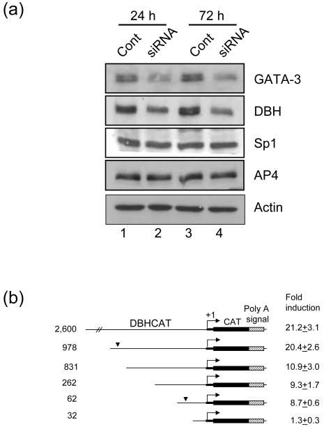
Fig. 2.
GATA-3 regulates expression of DBH. (a) The expression of DBH is reduced by GATA-3 siRNA. SK-N-BE(2)C cells were transfected with GATA-3 siRNA (lane 2, 4) or control siRNA (lane 1, 3) using Lipofectamine. After 24 (lane 1, 2) or 72 h (lane 3, 4) of transfection, cells were harvested and Western blot analyses were performed. The expression of GATA-3, DBH, Sp1, AP4 was detected with their specific antibodies as indicated. β-actin was detected as a loading control. (b) GATA-3 transactivation of serially deleted DBH promoter. HeLa cells were cotransfected with DBH-CAT reporter plasmids and pcDNA/GATA3 or empty vector at a molar ratio of 0.2. Fold induction by effector plasmid cotransfection is presented as mean ± SEM value from six to nine independent samples. The numbers on the left of the diagram represent the size of the DBH promoter. The bent arrow represents the DBH transcription start site. The bold thick line denotes the 5′ untranslated sequences and the thin line denotes the 5′ upstream sequences of the DBH gene. The arrowhead represents the GATA-3 response region of the DBH promoter.

