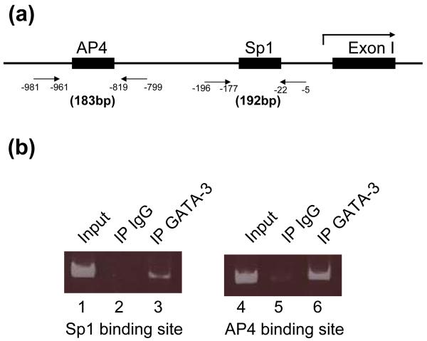
Fig. 7.
ChIP analysis indicates in vivo interaction of GATA-3 with SP1 and AP4 on the DBH promoter. (a) Schematic drawing of primers to detect Sp1 and AP4 binding sites by PCR. The first exon of human DBH gene is shown (Exon I). The numbers indicate nucleotide position in the DBH promoter. (b) The protein-DNA complexes were immunoprecipitated using antibodies against GATA-3 (lane 3, 6). As a negative control, rabbit IgG was used (lane 2, 5). Lane 1 and 4 show input DNAs. PCR was performed with primer sets shown (a) as described in experimental procedure. One twelfth (lanes 1, 4) or one third (lanes 2, 3, 5, 6) of the reaction PCR products were run in 7% polyacrylamide gel.

