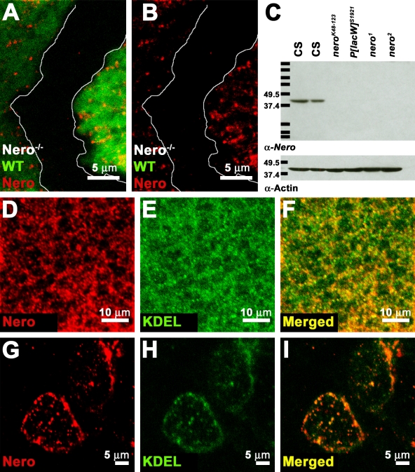
Figure 2.
Nero localizes to the ER. (A–C) Antibodies generated against Nero specifically detect the Nero protein on both immunohistochemical preparations and Western blots. (A and B) Nero antibody fails to recognize the protein in mutant clones marked by the absence of GFP (genotype: y w hs-FLP; FRT82B nero1/FRT82B Ubi-GFP). White lines mark the clonal boundary. WT, wild type. (C) The Nero antibody detects a single ∼39-kD band in Canton-S (CS) L2 larval protein extracts (first and second lanes) but fails to detect the protein in protein extracts from nerok48-123 (third lane), P[lacW]s1921 (fourth lane), nero1 (fifth lane), and nero2 (sixth lane) L2 larvae on Western blots. Protein lysates from 10 larvae were loaded in each lane except the second lane, in which only five larvae were loaded. Actin was probed as a loading control. Protein standards run along with larval lysates are marked with black bars. Their sizes are recorded in kilodaltons on the left. (D–F) Double labeling of Canton-S third instar wing discs using the Nero antibody and the KDEL antibody shows extensive colocalization, demonstrating that Nero is ER associated. (G–I) Double labeling of Canton-S third instar garland cells using Nero and KDEL antibodies.

