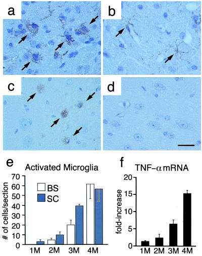
Figure 2.
Microglia activation and expansion in Sandhoff disease mice. Immunostaining with anti-F4/80 antibody shows amoeboid microglia in the brainstem of 4-month-old Sandhoff disease mouse (a) and ramified microglia in the brainstem of age-matched control mouse (b). Staining with GSIB4 shows amoeboid microglia in the spinal cord of a 4-month-old Sandhoff disease mouse (c) but not in a control mouse (d). (Bar = 20 μm.) (e) Quantitation of GSIB4-positive microglia in brainstem (BS) and spinal cord (SC) of Sandhoff mice. The GSIB4-positive cells were counted in semisequential sections of brain and spinal cord. Data are mean ± SEM (n = 3–4). (f) TNF-α mRNA expression levels in the spinal cord of Sandhoff disease mice compared with age-matched control mice. (SD). Data are mean ± SEM (n = 3).

