
Fig. 3.
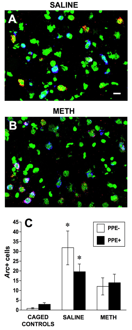
Confocal images showing Arc mRNA (cy-3/red), preproenkephalin mRNA (PPE, cy-5/blue) and nuclear (SYTOX Green) staining in the dorsomedial striatum of a saline- (4 × 1 mL/kg, s.c. at 2-hr intervals; A) and a methamphetamine- (METH; 4 × 10 mg/kg, s.c. at 2-hr intervals; B) pretreated rat. The average number (± SEM, n= 4 for caged controls and n=7 for both saline- and METH-pretreated groups) of PPE− and PPE+ neurons expressing Arc mRNA in the cytoplasm in a 0.7 mm × 0.7 mm field of the dorsomedial striatum (C). * Significantly different from respective phenotype in caged controls, p<0.05. Scale bar, 10 µm.