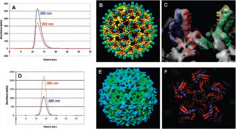
Figure 3.
Characterization of the HBV (A-C) and Qβ (D-F) capsids containing 1. (A and D) Size exclusion chromatography (Superose-6), with retention times matching those of intact native capsids. (B and E) Cryo-electron microscopy image reconstructions. (C and F) X-ray crystal structures docked into the cryo electron density maps. Characterization data for Qβ incorporating 2 may be found in Supporting Information.

