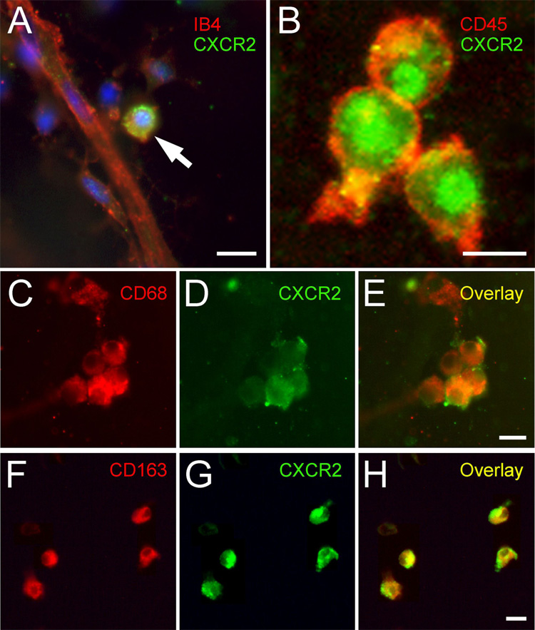
FIGURE 3.
Immunocytochemical localization of CXCR2 in aortic cultures. A: Immunofluorescent staining of angiogenic outgrowth from cultured rat aortic ring shows expression of CXCR2 (green) in a round cell (arrow) next to a neovessel highlighted with the endothelial marker IB4 (red); nuclei are stained with DAPI (blue). B: Confocal image shows coexpression of CXCR2 (green, perinuclear staining) with the leukocyte marker CD45 (red) in small cluster of round cells found close to the roots of the angiogenic outgrowth, near the aortic explant. C–H: Double immunofluorescent staining of aortic cultures for CD68 (C, red) or CD163 (F, red) and CXCR2 (D, G green), demonstrates co-localization of CXCR2 in macrophages (overlays in E and H). Scale bars = 10 µm.

