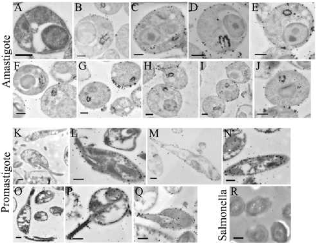
Fig. 5.
Localization of MSP in stationary phase LcJ promastigotes or axenic amastigotes using immunogold staining and electron microscopy. LcJ amastigotes (panels A–J) or promastigotes (panels K–Q), or Salmonella typhimurium (panel R) were fixed and embedded in LR White resins. Thin sections were incubated with buffer alone (A, K) or with affinity-purified anti-MSP antiserum (panels B–J, L–Q, R) followed by secondary antibody conjugated to ultra-small gold particles (< 0.8 nm). Scale bars represent 0.5 µm in panels A–Q, or 0.1 µm in panel R.

