
Fig. 4.
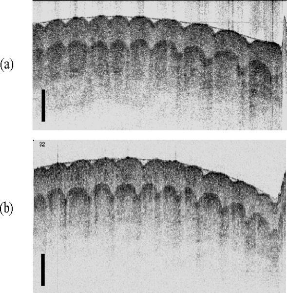
(a) Image of a human finger acquired in vivo with the SD-OCT system at 38 fps (256 axial × 500 transverse pixels, 2.1 × 5.0 mm). (b) Image of the same human finger (250 axial × 500 transverse pixels, 2.5 × 5.0 mm) acquired at 4 fps using a state-of-the-art TD-OCT system. The scale bars represent 0.5 mm.