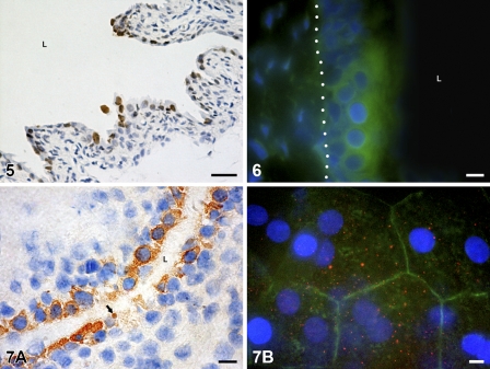
Figure 7.
Immunodetection of active caspase-3 in the urothelium. (A) Brown color shows positive reaction in the cytoplasm of the majority of urothelial cells and in one apoptotic body (arrow) in the tissue section on postnatal day 7. Nuclei are stained with hematoxylin. L, lumen of the urinary bladder. (B) Positive reaction is manifested as red dots (red fluorescence) in the cytoplasm of superficial cells. Actin filaments (green fluorescence) are arranged as a cortical belt close to the cell borders. Bird’s-eye view of the luminal urothelial surface on the tissue piece, postnatal day 10. Nuclei are stained with DAPI. Bar = 10 μm.

