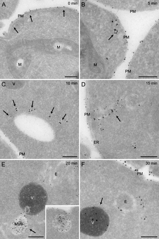
Figure 2.
Time-course labeling of the yeast endocytic compartments with positively charged Nanogold. Spheroplasts obtained from the wild-type SEY6210 strain were incubated in the presence of 4 nmol of positively charged Nanogold at 4C for 15 min before being transferred to room temperature. Aliquots of the resuspension were collected after 0 (A), 5 (B), 10 (C), 15 (D), 20 (E), and 30 min (F) before being fixed and processed as described in Materials and Methods. Arrows point to the membranous structures that become labeled at the indicated times, e.g., PM (A), endocytic vesicles (B), cluster of early endosome (EE) vesicles (C), EE (D), MVB (E), and vacuole (F). (E) inset, MVB with clearly resolved internal vesicles. The Nanogold uptake times are indicated at the top of each panel. E, endosomes; M, mitochondria; MVB, multivesicular body; PM, plasma membrane; V, vacuole. Bar = 200 μm.

