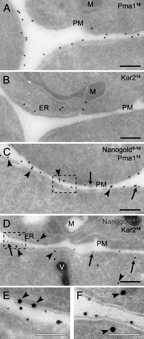
Figure 4.
Double labeling of cryosections with positively charged Nanogold particles and immunological reactions. Wild-type cells (SEY6210) grown to log phase were converted to spheroplasts and incubated with 4 nmol of positively charged Nanogold at 4C for 15 min before being immediately fixed. Cryosections with (C,D) or without (A,B) a silver enhancement reaction were incubated with anti-Pma1 (A,C) or anti-Kar2 (B,D) antibodies, followed by 15-nm protein A–gold particle conjugates. Arrows indicate Nanogold particles, whereas arrowheads indicate immunogold labeling. (E,F) Enlargements of defined labeled areas of C and D, respectively, to better illustrate the clear difference in size and density between the silver-enhanced Nanogold and gold conjugated to antibodies. The size of the particles is indicated at the top of each panel. ER, endoplasmic reticulum; M, mitochondria; PM, plasma membrane; V, vacuole. Bars: A–D = 200 nm; E,F = 100 nm.

