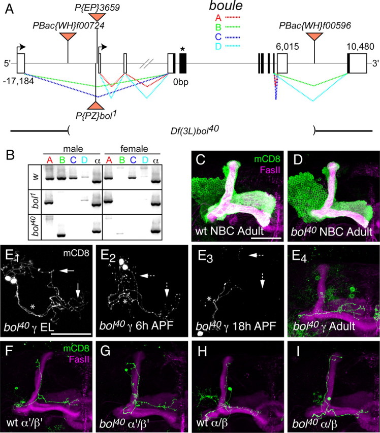
Figure 7.

Loss-of-function analysis of Boule in MB neurons. A, Schematic of boule gene illustrating alternative splicing of exons and transposon insertions. The boule gene is predicted to make four different transcripts through alternative splicing and alternative promoter use. The two promoters are represented by black arrows. EP3659 is inserted upstream of the second transcriptional start site in the same orientation as boule. The bol1 P-element insertion is located just upstream of EP3659, but in the opposite orientation. Flippase-mediated recombination between two piggyBac elements containing FRT sites (PBac{WH}f00724 and PBac{WH}f00596) was used to create a small deficiency in boule (Df(3L)bol40), which deletes the majority of the coding sequence (see Materials and Methods). The coding sequence is shown in black, and the asterisk denotes the location of the RNA-binding domain. The dashed lines represent alternative splicing patterns with colors corresponding to individual splicing isoforms. B, RT-PCR of boule transcript expression in w1118 (wt control), bol1, and bol40 adult male and female flies. wt males express all four boule transcripts, whereas females do not express bol-B. bol1 homozygous mutant flies do not express bol-B or bol-C. No full-length transcripts are detected in bol40 homozygous mutant males or females. A truncated transcript of bol-B, most likely corresponding to the splicing of exon 1 to exon 11, is expressed in bol40 males. RT-PCR for ubiquitously expressed α-tubulin (α) mRNA is used as a positive control for RT-PCR. C, D, MARCM NBCs for wt control MB neurons (C) and bol40 mutant MB neurons (D) have grossly similar neuronal morphology. E, Developmental time course analysis of γ neuron pruning in bol40 mutant single-cell or two-cell MARCM clones. As previously described for wt MB γ neurons (Watts et al., 2003), bol40 mutant γ neurons have intact dorsal and medial axon branches in the third-instar larva (E1) and show initial signs of axon fragmentation at 6 h APF (E2). By 18 h APF (E3), the axon branches have degenerated back to the primary axon [marked with an asterisk (*)] and only the medial branch is reextended in the adult (E4). The dashed arrows denote degenerating axons. F–I, Single-cell MARCM clones of wt and bol40 mutant α′/β′ (F, G, respectively) and α/β (H, I, respectively) showing normal morphology in the adult. All panels show confocal Z projections of the MB neurons. n ≥ 12 brains for each experiment. Scale bars, 50 μm.
