
Figure 7.
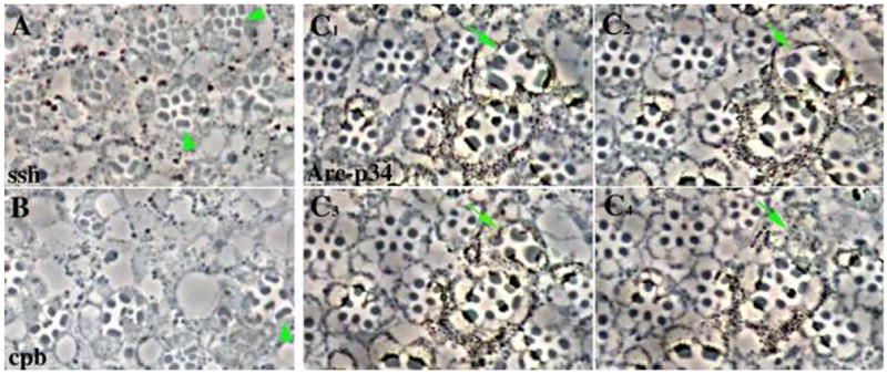
Widened rhabdomeres are found in photoreceptor cells mutant for genes that are involved in the reorganization of the actin cytoskeleton. The FLP/FRT system was used to make somatic mosaics of the mutant alleles of (A) ssh1–11, (B) cpbM143, and (C1–C4) Arc-p34KG04658. The mosaic eyes were fixed and sectioned. (A, B) Examples of widened or split rhabdomeres are indicated by arrowheads. (C1–C4) contain a series of four adjacent serial sections. The arrow points to an ommatidia containing photoreceptors with widened rhabdomeres. These rhabdomeres do not extend the full length of the retina and terminate within the serial sections shown. The Arc- p34KG04658 allele is marked with a mini-white gene. Heterozygous cells have low levels of pigment, with the homozygous mutant cells being more darkly pigmented.