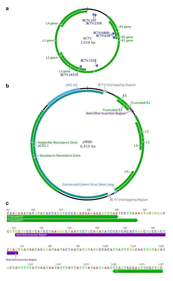
Figure 1.

Map of the pWSRi plasmid and its construction. (a) The circular map of the BCTV genome displays the positions of the primers (blue arrows) used to construct pWSRi relative to the positions of the viral genes (green arrows). (b) The circular BCTV genome was inserted into a pCR2.1 plasmid vector (indicated in blue). In plant cells, the viral vector is released by recombination within the overlap (repeated) regions of the viral genome marked in red. The full-length BCTV L1, L2, L3, L4, and R3 are indicated with green arrows. The positions of the truncated R1 and R2 genes, interrupted by the NotI-XbaI linker for insertion of targeting sequences (in purple), are also indicated in green. Direction of the arrows indicates direction of gene transcription. (c) The nucleotide sequence near the targeting sequence insertion site is shown. The truncated R1 and R2 reading frames are denoted by arrows underneath the sequences; 14 nt were deleted from the 3' end of R2 and 626 nt were deleted from within R1. The remainder of the R1 transcription unit continues downstream of the XhoI site. The L3 reading frame, which is not truncated, is similarly marked with an arrow in the direction of the transcription. The linker containing NotI/XhoI cloning sites is shown in purple.
