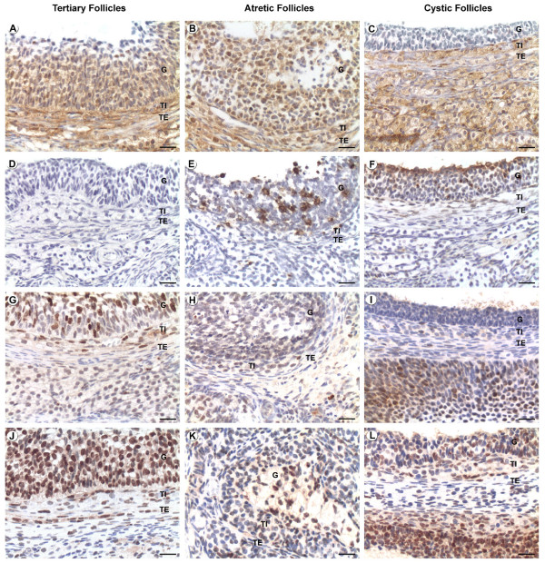
Figure 1.
Localization of caspase-3, Ki-67 and PCNA by immunohistochemistry and in situ apoptosis by TUNEL. Positive staining is shown as brown colouring of the cytoplasm/nucleus of the cells. Figures A, D, G and J correspond to healthy tertiary follicles; B, E, H and K correspond to atretic follicles and C, F, I and L correspond to cystic follicles. (A-C) caspase-3 immunolocalization, (D-F) TUNEL, (G-I) Ki-67 immunolocalization and (J-L) PCNA immunolocalization. G: Granulosa, TI: Theca Interna, TE: Theca Externa. Bars = 20 μm.

