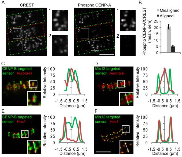
Figure 1.
Phosphorylation of a kinetochore Aurora B substrate depends on the microtubule attachment state. (A-B) Hela cells with both incorrect (green boxes, A) and bi-oriented (red boxes, A) attachments were fixed and stained for kinetochores (CREST) and phospho-CENP-A. Insets show stronger phospho-CENP-A staining on unaligned (1) vs. aligned (2) kinetochores. The phospho-CENP-A/CREST ratio was calculated at individual aligned (N=146) and unaligned (N=89) kinetochores from multiple cells (B). (C-F) Hela cells expressing either the CENP-B-targeted (C,E) or Mis12-targeted (D,F) sensor were fixed and stained for either Aurora B (C,D) or Hec1 (E,F) as markers for the inner centromere and outer kinetochore, respectively. YFP emission (green) shows sensor localization relative to Aurora B or Hec1 (red). Insets show individual centromere pairs used for linescans. Scale bars 5 μm.

