Figure 3.
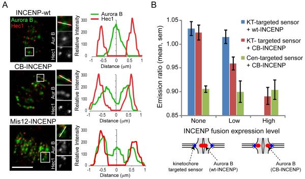
Positioning Aurora B closer to the kinetochore leads to increased phosphorylation of kinetochore substrates. (A) U2OS cells expressing the indicated vsv-tagged INCENP fusion proteins were fixed and stained for Aurora B (green) and Hec1 (red). Scale bars 5 μm. (B) Hela cells were transfected with a phosphorylation sensor together with mCherry-tagged wt-INCENP or CB-INCENP as indicated, imaged live, and the YFP/TFP emission ratio was calculated. Cells were grouped based on levels of the INCENP fusion proteins at centromeres, calculated based on mCherry fluorescence intensity (wt-INCENP was not present at as high levels as CB-INCENP). Each bar represents an average over multiple cells, ≥30 kinetochores analyzed per cell.

