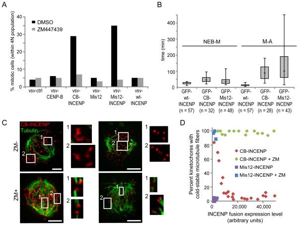
Figure. 4.
Positioning Aurora B closer to the kinetochore activates the spindle checkpoint and destabilizes kinetochore-microtubule attachments. (A) U2OS cells expressing the indicated proteins were released from a G1/S block with or without ZM447439 for 14 hrs. The mitotic index was determined by propidium iodide/MPM2 mAb labeling and FACS analysis. One representative experiment out of 3 is shown. (B) Box-and-whisker plots (median, interquartile range, full range) showing time from nuclear envelope breakdown to metaphase (NEB-M) and from metaphase to anaphase (M-A) of U2OS cells expressing the indicated INCENP fusion proteins. (C-D) Hela cells expressing vsv-tagged CB-INCENP or Mis12-INCENP were treated with or without ZM447439, then analyzed for cold-stable microtubules (green) and vsv immunofluorescence (red). Brightness of vsv-Mis12-INCENP staining is enhanced relative to vsv-CB-INCENP so that it is visible. The percent of kinetochores with cold-stable microtubules was determined and plotted vs. expression level of the INCENP fusion proteins (D). Each data point represents >80 kinetochores from one cell.

