
Figure 3.
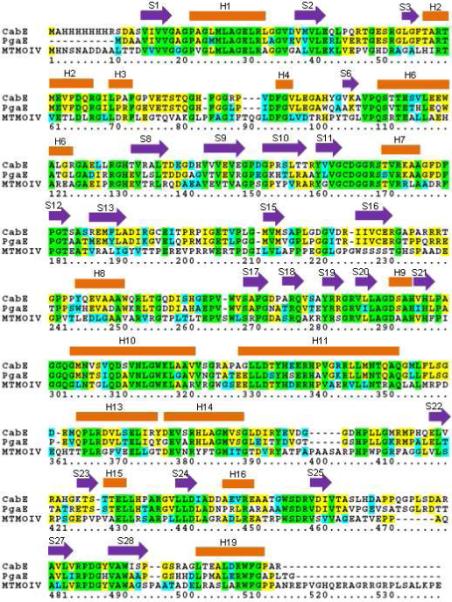
Sequence alignment of MtmOIV, PgaE, and CabE. Green shading indicates residues that are completely conserved, yellow indicates partially conserved, and cyan indicates similar residues. Secondary α-helix structures are labeled as orange blocks above the residues, and β-sheets with purple arrows. Structural labels have the same numbering as the PgaE structure (44).