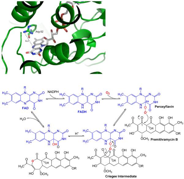
Figure 8.
A. Left: View of the catalytically important arginine-52 (R52, green stick, N = blue, O = red) residue of MtmOIV above the flavin ring of FAD (gray stick, N = blue, O = red, P = orange). The measured distance (dotted line) between the N-atoms of the guanidine residue of R52 and the flavin ring surface is ∼ 6.2 Å in the shown conformation. The remainder of the enzyme (ribbon) is depicted in green.
B. Below: Suggested Baeyer-Villiger reaction mechanism of the MtmOIV reaction, involving the peroxyflavin and Criegee intermediates. R = deoxysugar chains

