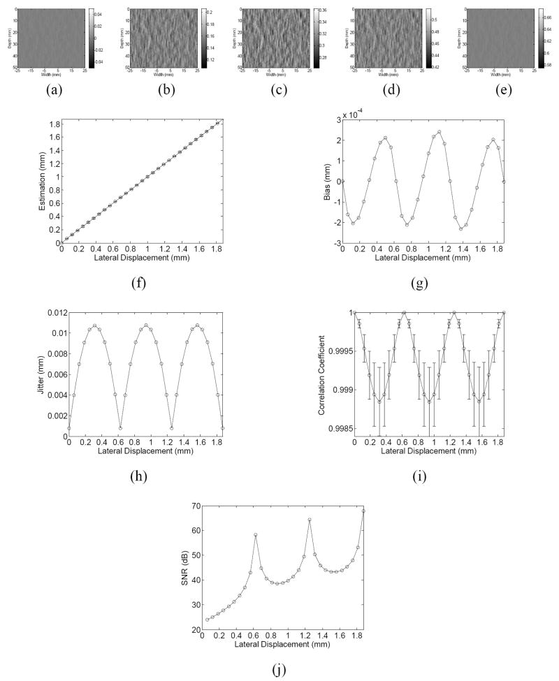
Figure 2.
The estimated lateral displacement images when the applied lateral displacement is equal to (a) 0, (b) 0.25, (c) 0.5, (d) 0.75 or (e) 1 pitch, and (f) average of the estimated displacement, (g) the bias and (h) jitter of estimation, (i) average correlation coefficient and (j) signal-to-noise (SNR) ratio of lateral displacement estimation as a function of the applied lateral displacement ranging from 0 to 3 pitch. In (a)-(e), the gray scale is equal to the true displacement ± 0.05 mm. In (f) and (i), the error bar represent one standard deviation (SD). (pitch = 0.625 mm, beamwidth = 2 mm).

