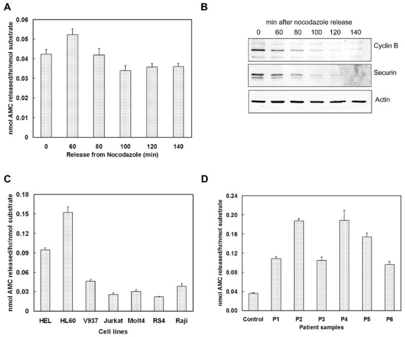
Figure 4.

Determination of Separase activity in cells and leukemia samples. (A). Separase activity during mitotic progression. HeLa cells were synchronized with double thymidine and arrested at G2/M with nocodazole. Metaphase arrested cells were harvested for lysis at 0, 60, 80, 100, 120 and 140min after release from nocodazole block. Separase activity assay was performed with 20μg protein from each time point. Each time point is mean ± SEM of three observations. (B). Immunoblotting of Cyclin B and Securin levels in HeLa cells after release from nocodazole arrest. (C). Separase activity assay in a panel of leukemic cell lines. 20μg total protein from each line was analyzed. (D). Separase activity in pediatric AML samples. Peripheral blood cells from normal individual were used as control. 25μg total protein for each sample was analyzed.
