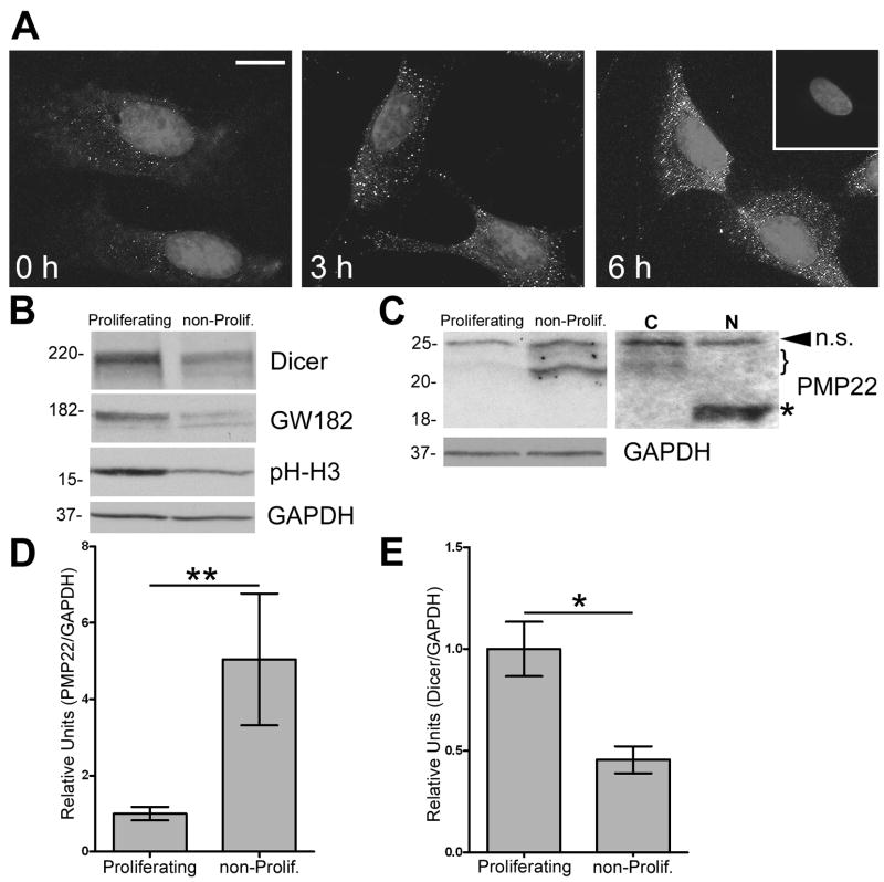
Figure 1. GW body formation and Dicer expression are enhanced in actively-proliferating Schwann cells.
(A) Rat Schwann cells were subjected to growth arrest, followed by the addition of serum (for time-points indicated) to induce cellular growth. Cells were fixed and labeled with a human anti-GWB antibody. Hoechst dye is used to visualize nuclei. The inset in the upper right corner represents a no primary antibody control. Scale bar, 10 μm. (B) Western blot analysis on total lysates (40 μg/lane) of proliferating and non-proliferating (non-prolif.) cells are shown using the indicated antibodies. Phospho-Histone H3 (pH-H3) serves as a mitotic marker. (C) On the anti-PMP22 Western blot, the arrowhead indicates a non-specific (n.s.) band whereas the bracket denotes differentially glycosylated isoforms of PMP22. Upon incubation of the cell lysates with PNGase F (N), the indicated ~22 kDa PMP22 bands shift to the core 18 kDa core protein (*). C: no enzyme control. (B, C) GAPDH serves as a loading control. Molecular mass in kDa. Quantification of three independent experiments reveals increased PMP22 (D) and decreased Dicer (E) expression in non-proliferating, as compared to proliferating cells (**p<0.01; *p<0.05). (D, E) Values are expressed as relative units normalized for GAPDH.

